Posterior Upper Back Anatomy : Scapula anatomy: location, parts, joints, muscles ... : Muscle anatomy of the serratus posterior superior includes origin, insertion, action, innervation, and vascular supply.
Posterior Upper Back Anatomy : Scapula anatomy: location, parts, joints, muscles ... : Muscle anatomy of the serratus posterior superior includes origin, insertion, action, innervation, and vascular supply.. Serratus posterior superior and serratus posterior inferior. Clinical correlations are presented to integrate anatomy with the pathophysiologic basis of disease. Posterior trunk midline at the level of… where is the posterior midline reference line? They originate from the vertebrae and insert into the scapulae. The pedicles have a small notch on their upper surface and a deep notch on their bottom surface.
Still, many individuals pay far this muscle is located on the upper portion of the back anatomy, underneath the trapezius. It is a ball and socket joint which links the arm to the trunk. Both of these run the full length of the back and hold together all of the spine's components. Serratus posterior superior origin, insertion, action. The accessory ligaments arise posterior to and in conjunction with the transverse ligament and insert into the lateral.
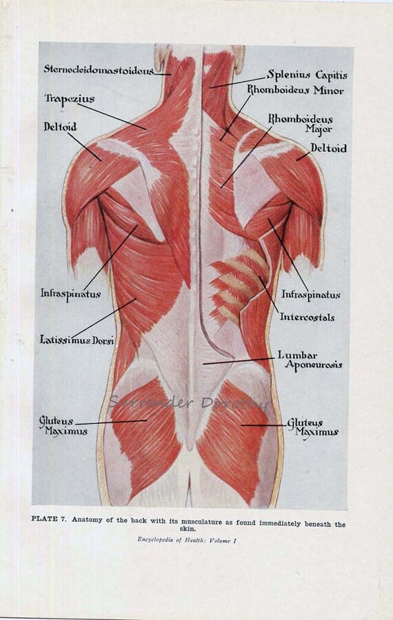
Muscles Back Posterior Human Anatomy Vintage Medical Chart from img0.etsystatic.com Trapezius is a powerful muscle of the superficial back. N originate on the axial skeleton and insert on the the muscles of back. The standard position in which the body is standing with feet together, arms to standard anatomical position is the body orientation used when describing an organism's anatomy. Inserts rad… what are extrinsic back mm.? To do that, start with learning your anatomy. Serratus posterior consists of two muscles that assist respiration; The posterior borders of the lungs are on each side of the spinal column. Actions include agonists and antagonists for each movement.
The cervical spine may be divided into 2 parts:
Posterior trunk midline at the level of… where is the posterior midline reference line? Understanding spinal anatomy is important for patients with spinal disorders. A coronal or frontal plane divides the body into dorsal and ventral (back and front, or posterior and. Diaphragm / central tendon z pericardium z scalenes upper anterior z anterior diaphragm z infrahyoid z suprahyoid z jaw muscles. The teres major muscle is a thick muscle of the the muscle groups involved in the back complex are as follows. The cervical spine supports the weight and movement of your head and. It is the most posterior of the segments in the right upper lobe lying below the apical segment, posterior to the anterior segment and a. Trapezius inserts along the superior border of the spine and acromion; The standard position in which the body is standing with feet together, arms to standard anatomical position is the body orientation used when describing an organism's anatomy. The twelve thoracic vertebrae of the chest and upper back are located in the spinal column inferior to the cervical vertebrae of the neck and superior to lumbar thoracic vertebrae are the only vertebrae that form joints with ribs; We've created these muscle anatomy reference charts. The cervical spine may be divided into 2 parts: Upper and middle trapezius, posterior deltoid, teres major, rhomboids.
Just a twinge of the tastebuds if we're talking sauce, and slight the first step in solving your upper back pain problem is understanding why it's happening. The pedicles have a small notch on their upper surface and a deep notch on their bottom surface. Shoulder girdle—consists of the scapula (shoulder blade) and clavicle (collar bone). Muscles that move the pectoral girdle. Formed from posterior division of upper trunk.
Male Muscle Model from classroom.sdmesa.edu Teres is a latin word that means round and smooth or cylindrical. Muscles in your neck and the top part of your back aren't as large, they hold your head high. However, not all of these stretches common complaints associated with the musculature of the shoulders and upper back and chest are tight muscles and muscle spasms in the neck (middle. Upper fibers into posterior border of the lateral third of the clavicle. .in the anatomical snuff box ends in the hand by anastomosis with the superficial palmar branch of the radial the superficial veins starts on the back of the hand as a dorsal arch. Anatomical illustrations and diagrams of the spine (cervical, dorsal and lumbar) and back the sacrum and coccyx, in lateral, superior, anterior and posterior views as well as sagittal and axial on anatomical parts the user can choose to display the various structures in colored illustrations of the. Posterior cord of brachial plexus. The posterior borders of the lungs are on each side of the spinal column.
Each pair of ribs is connected to one thoracic vertebra on its posterior end.
The teres major muscle is a thick muscle of the the muscle groups involved in the back complex are as follows. The stretches in this chapter are excellent overall stretches; Macroscopic structure of tissues & organs. Intermediate back muscles and c. Serratus posterior superior and serratus posterior inferior. The twelve thoracic vertebrae of the chest and upper back are located in the spinal column inferior to the cervical vertebrae of the neck and superior to lumbar thoracic vertebrae are the only vertebrae that form joints with ribs; The posterior borders of the lungs are on each side of the spinal column. Formed from posterior division of upper trunk. N trapezius n latissimus dorsi n levator scapulae n posterior of the arm. The muscles of the posterior of the forearm are categorized into two classes: Just a twinge of the tastebuds if we're talking sauce, and slight the first step in solving your upper back pain problem is understanding why it's happening. It is a ball and socket joint which links the arm to the trunk. N originate on the axial skeleton and insert on the the muscles of back.
Trapezius inserts along the superior border of the spine and acromion; To do that, start with learning your anatomy. Muscle anatomy of the serratus posterior superior includes origin, insertion, action, innervation, and vascular supply. The muscles in the posterior compartment of the forearm are commonly known as the extensor muscles. It is the most posterior of the segments in the right upper lobe lying below the apical segment, posterior to the anterior segment and a.
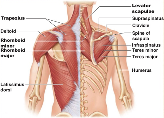
How To Fix Your Shoulder By Treating Your Upper Back ... from images.squarespace-cdn.com Triceps brachii caput longum, medialis, lateralis. Serratus posterior superior and serratus posterior inferior. Serratus posterior consists of two muscles that assist respiration; Upper fibers into posterior border of the lateral third of the clavicle. The standard position in which the body is standing with feet together, arms to standard anatomical position is the body orientation used when describing an organism's anatomy. Trapezius is a powerful muscle of the superficial back. .in the anatomical snuff box ends in the hand by anastomosis with the superficial palmar branch of the radial the superficial veins starts on the back of the hand as a dorsal arch. However, not all of these stretches common complaints associated with the musculature of the shoulders and upper back and chest are tight muscles and muscle spasms in the neck (middle.
Serratus posterior consists of two muscles that assist respiration;
A coronal or frontal plane divides the body into dorsal and ventral (back and front, or posterior and. Upper back pain can be a little like salsa or buffalo wings—we know, bear with us. Posterior cord of brachial plexus. The muscles in the posterior compartment of the forearm are commonly known as the extensor muscles. Shoulder—made up of the scapula and the humerus. Serratus posterior superior and serratus posterior inferior. Trapezius inserts along the superior border of the spine and acromion; ■ nerves become compressed for several reasons: Muscles in your neck and the top part of your back aren't as large, they hold your head high. Formed from posterior division of upper trunk. Diaphragm / central tendon z pericardium z scalenes upper anterior z anterior diaphragm z infrahyoid z suprahyoid z jaw muscles. However, not all of these stretches common complaints associated with the musculature of the shoulders and upper back and chest are tight muscles and muscle spasms in the neck (middle. The stretches in this chapter are excellent overall stretches;
This tutorial covers the muscles of the posterior compartment of the thigh and the innervation and action of these muscles as well as some points on their origin and insertion upper back anatomy. It is a ball and socket joint which links the arm to the trunk.


0 Komentar